
Scientific Calendar January 2024
Diffuse large B cell lymphoma
How do abnormal lymphocytes react with WPC measurement channel reagents?
Membranes of abnormal lymphocytes are more readily permeated, causing higher fluorescence signals.
Abnormal lymphocytes shrink during the reagent treatment and subsequently show low fluorescence signals and smaller size.
During the reagent reaction, the abnormal lymphocytes expand to increase their volume.
Congratulations!
That's the correct answer!
Sorry! That´s not completely correct!
Please try again
Sorry! That's not the correct answer!
Please try again
Notice
Please select at least one answer
Scientific background
Diffuse large B cell lymphoma: a common type of lymphoma
Diffuse large B cell lymphoma (DLBCL) is the most common type of non-Hodgkin lymphoma, making up around 25% of cases [1]. It is also the sixth most common cause of death due to cancer [2]. DLBCL typically presents with abnormal lymphocytes that have large nuclei, basophilic cytoplasm and a high proliferation rate [3].
Flow cytometric immunophenotyping is commonly used to classify the disease.
Detection of abnormal lymphocytes with the XR-Series analyser
Abnormal lymphocytes have certain characteristics that make it possible to differentiate them from normal ‘healthy’ cells. A well-described trait of abnormal lymphocytes is an altered membrane composition with a high lipid content [4] and the fact that they have more apparent DNA when compared to normal lymphocytes, as they rapidly proliferate [5]. Sysmex XR-Series analysers take advantage of these special characteristics of the cell membranes in order to identify the cell, making use of specific channels and proprietary reagents.
In the WDF measurement channel, the lysis reagent reacts rather gently and leaves the internal structure of the cells largely intact while allowing the fluorescent marker to enter the cells and label primarily the RNA inside. Depending on the fluorescence signal of the cell clusters, specific flags are triggered, pre-classifying the sample into negative, reactive, potentially malignant or negative, and potentially malignant or reactive. Based on this pre-classification in the WDF measurement channel, the flags ‘Blasts/Abn Lympho?’ or the combination ‘Blasts/Abn Lympho?’ and ‘Atypical Lympho?’ are triggered. These lead to an automated reflex measurement in the WPC measurement channel.
The lysis reagent of the WPC measurement channel, on the other hand, has a stronger effect on the membrane lipids than the ones used in the WDF channel. Additionally, abnormal cells, due to their specific membrane composition and readily permeated membranes, allow greater access to their nuclear DNA [5], which is then stained by the specific WPC fluorescent marker. The higher the degree of membrane perforation, the more cellular content leaks through the pores, causing the cell to shrink. Therefore, abnormal lymphocytes will present with a smaller size signal and higher fluorescence signal [5]. Immature cells, such as blasts or haematopoietic progenitor cells, on the other hand, have a lower lipid content and are more resistant to lysis [4]. Therefore, they will give off lower fluorescence signals and remain larger.
Combining the information from both measurement channels, WDF and WPC on XR-Series analysers, makes it possible to differentiate between negative, reactive or suspected malignant cells [6].
Case and results interpretation
A 68-year-old patient with a main complaint of confusion and lethargy was admitted to the ICU. Haematology lab results revealed anaemia, leucocytopenia, and thrombocytopenia, as well as an abnormal white blood cell differential.
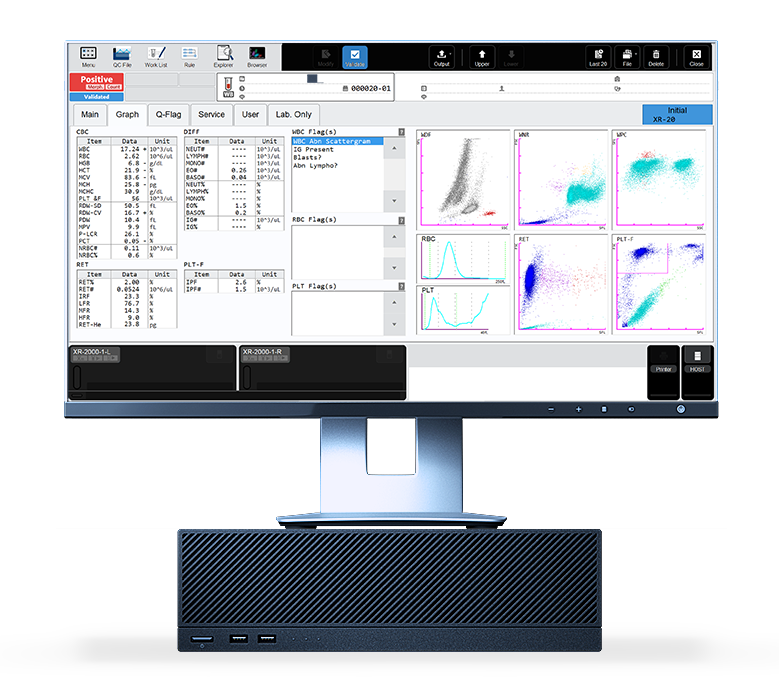
Fig. 2 The presence of abnormal lymphocytes can be observed in the WPC scattergram of the analyser.
The blood smear showed abnormal blast-like looking lymphocytes, some of which had a cleft nucleus.
Subsequent immunophenotyping results confirmed a suspected diagnosis of diffuse large B cell lymphoma. Additionally, the patient developed an infection with Streptococcus parasanguinis, which was confirmed by a blood culture.
Scientific background
Diffuse large B cell lymphoma: a common type of lymphoma
Diffuse large B cell lymphoma (DLBCL) is the most common type of non-Hodgkin lymphoma, making up around 25% of cases [1]. It is also the sixth most common cause of death due to cancer [2]. DLBCL typically presents with abnormal lymphocytes that have large nuclei, basophilic cytoplasm and a high proliferation rate [3].
Flow cytometric immunophenotyping is commonly used to classify the disease.
Detection of abnormal lymphocytes with the XR-Series analyser
Abnormal lymphocytes have certain characteristics that make it possible to differentiate them from normal ‘healthy’ cells. A well-described trait of abnormal lymphocytes is an altered membrane composition with a high lipid content [4] and the fact that they have more apparent DNA when compared to normal lymphocytes, as they rapidly proliferate [5]. Sysmex XR-Series analysers take advantage of these special characteristics of the cell membranes in order to identify the cell, making use of specific channels and proprietary reagents.
In the WDF measurement channel, the lysis reagent reacts rather gently and leaves the internal structure of the cells largely intact while allowing the fluorescent marker to enter the cells and label primarily the RNA inside. Depending on the fluorescence signal of the cell clusters, specific flags are triggered, pre-classifying the sample into negative, reactive, potentially malignant or negative, and potentially malignant or reactive. Based on this pre-classification in the WDF measurement channel, the flags ‘Blasts/Abn Lympho?’ or the combination ‘Blasts/Abn Lympho?’ and ‘Atypical Lympho?’ are triggered. These lead to an automated reflex measurement in the WPC measurement channel.
The lysis reagent of the WPC measurement channel, on the other hand, has a stronger effect on the membrane lipids than the ones used in the WDF channel. Additionally, abnormal cells, due to their specific membrane composition and readily permeated membranes, allow greater access to their nuclear DNA [5], which is then stained by the specific WPC fluorescent marker. The higher the degree of membrane perforation, the more cellular content leaks through the pores, causing the cell to shrink. Therefore, abnormal lymphocytes will present with a smaller size signal and higher fluorescence signal [5]. Immature cells, such as blasts or haematopoietic progenitor cells, on the other hand, have a lower lipid content and are more resistant to lysis [4]. Therefore, they will give off lower fluorescence signals and remain larger.
Combining the information from both measurement channels, WDF and WPC on XR-Series analysers, makes it possible to differentiate between negative, reactive or suspected malignant cells [6].
References
[1] Swerdlow SH et al. (2016): The 2016 revision of the World Health Organization classification of lymphoid neoplasms Blood. 127(20):2375-2390
[2] McNally GA et al. (2011): B-cell lymphoma, unclassifiable: a review of the literature. Clin J Oncol Nurs. 15(2):189-93.
[3] Padala SA et al. (2023): Diffuse Large B-Cell Lymphoma. Treasure Island (FL): StatPearls Publishing
[4] Gottfried EL et al. (1967): Lipids of human leukocytes: relation to celltype. J Lipid Res. 8(4):321-7.
[5] Kawauchi S et al. (2013): The Positions of Normal Leukocytes on the Scattergram of the Newly Developed Abnormal Celldetection Channel of the XN-Series Multi-parameter Automated Hematology Analyzers. Sysmex J Int; 23(1): 1
[6] Schuff-Werner P et al. (2016): Performance of the XN-2000 WPC channel-flagging to differentiate reactive and neoplastic leukocytosis. Clin Chem Lab Med; 54(9): 1503
Download our white paper

The fluorescence technology and channel-specific reagents of the XN-Series provide much more than just a cell count. Depending on their cell membrane composition, cells are perforated and labelled differently, disclosing information about the cells’ activity, maturity stage, and malignancy. This white paper sheds a light on how the XN measures cell functionality.
